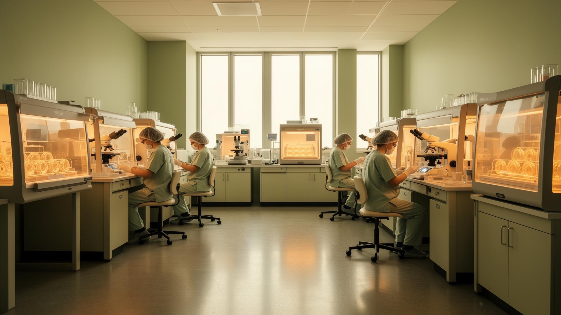
An embryo’s earliest days involve extraordinary changes, most of them invisible to the naked eye. In an IVF laboratory, a fertilised egg begins dividing, organising its cells and developing towards a stage that may allow transfer to the uterus. Time-lapse technology lets embryologists follow this development through images captured inside a specialised incubator. When patients ask me about Time-lapse embryology benefits, I explain that the value lies in understanding development more continuously—not in guaranteeing which embryo will become a baby.
In my practice, I help couples distinguish a useful laboratory tool from an essential treatment. Time-lapse imaging can add information to embryo assessment, but it does not replace skilled embryologists, careful clinical judgement or personalised fertility care.
What happens inside a time-lapse incubator?
During IVF, eggs are fertilised in the laboratory and the resulting embryos are cultured under carefully controlled conditions. With conventional assessment, embryologists usually examine embryos at selected intervals under a microscope. These observations provide important information about their appearance and developmental stage.
A time-lapse incubator combines a controlled culture environment with an integrated camera system. It takes images at regular intervals, creating a visual record of each embryo’s development. Embryologists can review this sequence without repeatedly removing the culture dish for routine visual checks.
The result is not simply a beautiful video. It shows when certain developmental events occur and how the embryo moves between stages. For example, an embryologist may observe whether cells divide in an orderly pattern or whether unusual divisions occur between standard observation times.
An embryo may eventually reach the blastocyst stage, when it has a fluid-filled cavity and groups of cells that will contribute to the baby and placenta. Not every fertilised egg reaches this stage, and time-lapse technology cannot prevent natural developmental arrest.
Understanding the benefits—and their limits
I find it helpful to separate what time-lapse systems clearly do from what patients understandably hope they will do.
Their practical advantages may include: - More continuous observation of development than occasional visual checks. - Less need to move embryos out of the incubator for routine assessment. - A record that embryologists can review, discuss and use for laboratory quality improvement. - Additional information to support decisions about which embryo to consider for transfer first.
A stable culture environment matters because embryos are sensitive to changes in temperature and other conditions. However, well-run conventional laboratories also use rigorous procedures to protect embryos. A time-lapse incubator alone does not tell us whether one laboratory provides better care than another.
The main clinical question is whether using this technology improves outcomes that matter most, particularly the chance of a live birth. Current evidence does not consistently show that time-lapse selection improves live-birth outcomes over good conventional assessment for everyone undergoing IVF. More information does not automatically mean a better outcome.
I therefore discuss it as a possible additional laboratory tool rather than a requirement for successful treatment. If it involves an extra charge, patients deserve a clear explanation of the expected benefit, the uncertainty and the alternatives.
How embryo selection works
Embryologists assess embryos using several clues. These include cell number, symmetry, developmental stage and fragmentation—small pieces of cellular material that can appear between cells. Time-lapse imaging adds information about developmental timing, sometimes called morphokinetics. In simple language, this means looking at how an embryo changes over time.
Some systems also use computer algorithms or artificial intelligence to rank embryos. These tools look for patterns in images and timing data. Their usefulness depends on how they were developed, tested and applied within a particular laboratory. A score should support professional judgement, not replace it.
Importantly, an attractive appearance or reassuring developmental pattern cannot confirm that an embryo has the expected number of chromosomes. Time-lapse imaging is not a genetic test.
PGT-A, or preimplantation genetic testing for aneuploidy, is a separate procedure that checks sampled embryo cells for chromosome-number abnormalities. It has its own limitations and is not appropriate for every patient. Time-lapse imaging neither replaces PGT-A nor makes genetic testing automatically necessary.
Similarly, ICSI—intracytoplasmic sperm injection, where a single sperm is injected into an egg—is a fertilisation technique, not an embryo-monitoring method. These technologies serve different purposes. I explain each recommendation separately so that patients understand why it may, or may not, belong in their treatment plan.
Where it fits into personalised, whole-person care
There is no single patient profile for whom time-lapse imaging is always necessary. Its relevance depends on the laboratory’s systems, available embryos, previous treatment history and the decisions the team needs to make. Having very few embryos, for example, may leave limited scope for ranking embryos, although developmental observations can still be informative.
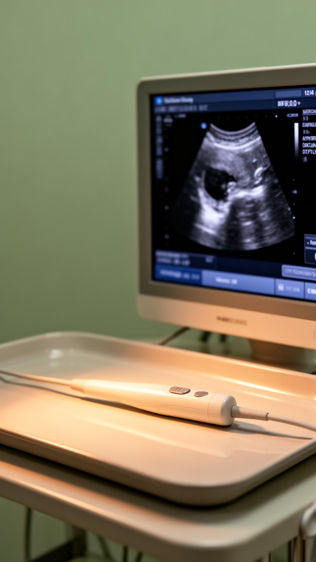
Before recommending treatment additions, I review the wider picture: age, ovarian response, sperm factors, uterine health and previous outcomes. Where indicated, a 3D ultrasound scan can help assess the shape of the uterine cavity. No imaging technology in the embryology laboratory can compensate for every clinical factor affecting implantation.
I encourage patients to ask: - How will these images change decisions in my case? - Is assessment performed by embryologists, software or both? - What evidence supports the system used by this laboratory? - Is this included in treatment, or is it an optional addition?
My 360-degree approach also includes preconception preparation, balanced nutrition, suitable physical activity and emotional support. Gentle yoga, breathing exercises and counselling may help patients manage stress and feel more supported during treatment. They should not be presented as ways to make an embryo implant, and unsuccessful treatment is never proof that someone failed to relax.
For patients interested in naturopathy, I prioritise safety. Supplements and herbal products need review because some may interfere with fertility medicines or be unsuitable around conception. Supportive wellness care should complement evidence-based treatment, not delay or replace it.
Key takeaways
Time-lapse embryology offers a detailed window into early embryo development and can support laboratory assessment while reducing routine handling. It cannot guarantee pregnancy, establish an embryo’s chromosome status or overcome every cause of infertility. I place the greatest value on an experienced laboratory team, transparent discussions about evidence and personalised protocols. Combined with thoughtful preconception care and compassionate support, technology should help us make informed decisions—not create pressure to choose every available add-on.
To schedule your personalized consultation with Dr. Munawwer Sana at Apollo Fertility in Kondapur, Hyderabad, or to arrange an online telehealth video consultation, click here to book your appointment.
Want a personalised plan based on your reports?
Book an online consultation with Dr. Sana, or see her at Apollo Fertility, Kondapur (Mon, Tue, Wed, Thu & Sat, 1:00 PM – 6:00 PM). No referral required.
References & clinical guidelines
- Indian Council of Medical Research — National ART Guidelines and the ART (Regulation) Act, 2021
- ESHRE — European Society of Human Reproduction and Embryology, clinical practice guidelines
- NICE CG156 — Fertility problems: assessment and treatment
- Royal College of Obstetricians and Gynaecologists — patient information and guidelines